ImageJ/FIJI is a powerful, easy-to-use, open-source image analysis software suite for 2D/3D scientific images. This hands-on course will provide a comprehensive understanding of image processing, analysis and quantification using Fiji. It is designed for scientists working with biological or biomedical image data.
Course details
This 3-day hands-on course will provide an in-depth introduction to bioimage analysis with Fiji. Participants will learn core features of the software as well as fundamental concepts of image processing and analysis. In addition, we will cover useful tools tailored for cell biology applications, and finish with an introduction to ImageJ macro writing for automating your analysis.
The course is intended for PhD students interested in image processing and analysis, particularly those working with microscopy data in biological and medical contexts. No prior experience with ImageJ/Fiji and image analysis knowledge is required. You will need to bring a laptop.
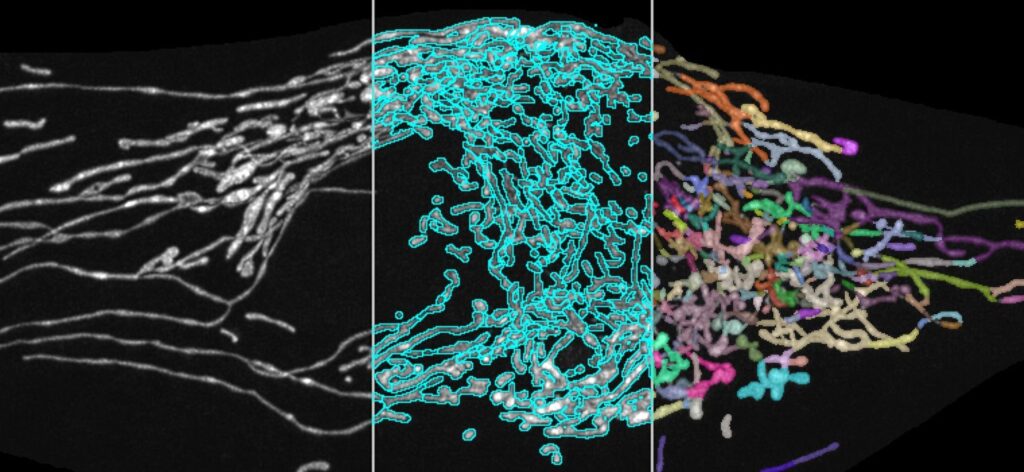
Key topics • Getting started with Fiji (installation, opening microscopy format images, plugins) • Image basics (pixels, image types, histogram, colors, stacks) • Creating multi-panel, publication-ready figures • Image processing (background subtraction, feature enhancement) • 3D visualization • Quantification (segmentation, cell counting intensity measurements) • Machine learning tools in Fiji • Automating your analysis with ImageJ macros • Work on your own data (if time permits)
By the end of this course, you will have a broad understanding of ImageJ/Fiji’s capabilities, empowering you to tackle a wide range of image analysis tasks in your research.
Meet the course organizers
Rolf Harkes
Post-doc at the Computational Pathology group. Imaging Scientist at the BioImaging Facility.
What I found most valuable about the OOA imageJ course was learning how to write macros on imageJ for analyzing the images, which is useful when you want to analyze many images in the same way. I would definitely recommend this course to others because even though I worked with imageJ before, I learned many new things.
The OOA ImageJ/Fiji course gave me an excellent, well-rounded overview of basic image analysis techniques, from generating publication-ready figures to writing macros, all of which I will apply to my work going forward. I would recommend this course to anyone who wants to learn the basics, brush up on their current knowledge, or to simply increase their confidence in using ImageJ
I highly recommend the Fiji course to both early and late-stage PhD students. It provides a solid foundation in image analysis, especially through macro writing. The organizers are friendly and always willing to help with any image analysis challenges during or after the course.
- The Fiji/ImageJ course has been incredibly valuable for learning how to perform various advanced image analysis techniques and automate them using macros. The instructors were very skilled and incredibly nice and helpful. I would highly recommend this course to others who are working with imaging data or planning to do so —it’s a great opportunity to deepen your understanding of image analysis and discover new ways to work efficiently.